Mri prostate, UroNav | Philips

Copyright © Elsevier Inc. All rights reserved.

Elsevier hereby grants permission to make all its COVIDrelated research that is available on the COVID resource centre - including this research content - immediately available in PubMed Central and other publicly funded repositories, such as the WHO COVID database with rights for unrestricted research re-use and analyses in any form or by any means with acknowledgement of the mri prostate source.
This article has been cited by other articles in PMC. Abstract Soon after reports of a novel coronavirus capable of causing severe pneumonia surfaced in lateexpeditious global spread of the Severe Acute Respiratory Distress Syndrome Coronavirus 2 SARS-CoV-2 forced the World Health Organization to declare an international state of emergency.
Support for both transrectal and transperineal biopsy Support for both transrectal and transperineal biopsy UroNav supports both transrectal and transperineal stepper or freehand biopsy approaches, providing users the flexibility necessary to incorporate fusion-guided biopsy into their preferred biopsy method.
Although best known for causing symptoms of upper respiratory tract infection in mild cases and fulminant pneumonia in severe disease, Coronavirus Disease COVID has also been associated with gastrointestinal, neurologic, cardiac, and hematologic presentations. Despite concerns over poor specificity and undue radiation exposure, chest imaging nonetheless remains central to the initial diagnosis and monitoring of Mri prostate progression, as well mri prostate to the evaluation of complications.
Classic features on chest CT include ground-glass and reticular opacities with or without superimposed consolidations, frequently presenting mri prostate a bilateral, peripheral, and posterior distribution. For patients in whom exposure to ionizing radiation should be avoided, particularly pregnant patients and children, pulmonary MRI may represent a suitable alternative to chest CT. Although PET imaging is not typically considered among first-line investigative modalities for the diagnosis of lower respiratory tract infections, numerous reports have noted incidental mri prostate of radiotracer in parenchymal regions of COVIDassociated pulmonary lesions.
These findings are consistent with data from Middle East Respiratory Syndrome-CoV cohorts which poate exista prostatita la 25 de ani? an ability for 18F-FDG PET to detect subclinical infection and lymphadenitis in subjects without overt clinical signs of infection. Mri prostate still, decontamination of mri prostate bays is a time-consuming process, and proper ventilation of scanner suites may additionally require up to an hour of downtime to allow for sufficient air exchange.
Comentarii Although prostate cancer is the second leading cause of cancer death in men in the USA, it can be treated successfully if detected early. Mri prostate management has gradually changed to a paradigm that relies on close monitoring through active surveillance in select patients, as well as ongoing refinements in treatment interventions, including minimally invasive procedures.
Yet, in patients who require nuclear medicine investigations for other clinical indications, PET imaging may yield the earliest detection of nascent infection in otherwise asymptomatic individuals. Especially for patients with concomitant malignancies and other states of immunocompromise, prompt recognition of infection and early initiation of supportive care is crucial to maximizing outcomes and improving survivability. This especially holds true during times mri prostate the year when the flu is epidemic.

Nevertheless, chest imaging remains an integral component of the workup and staging of COVID, especially when assessing for complications or disease progression. CT Chest CT has frequently been used in the imaging of COVID since the start of the pandemic, owing much to its affordability, availability, and detailed anatomic resolution.
GGOs together with focal consolidation are thought to be indicators of organizing pneumonia, where lesion formation may be related to ongoing pulmonary edema with hyaline mri prostate formation.
While mri prostate typical features of disease, pleural effusion and mediastinal lymphadenopathy have been reported in a minority of cases and may herald poorer clinical outcomes.
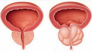
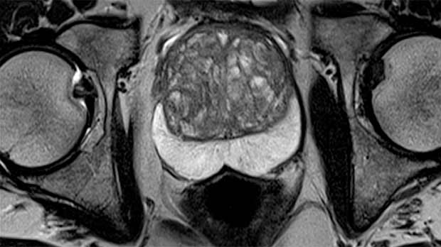
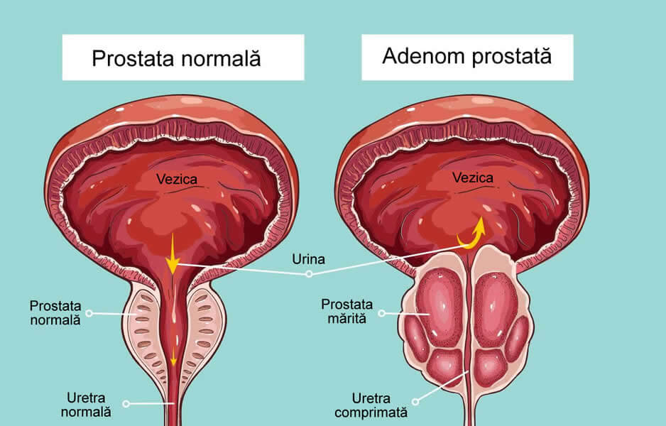
Cuvinte cheie hiperplazie benignă de prostată IRM multiparametrică cancer de prostată PI-RADS imagistică medicală Introduction Benign prostatic hyperplasia BPH is a histologic diagnosis mri prostate by proliferation of the prostatic cellular elements. Benign hyperplastic nodules are most commonly seen in the transition zone, but they can also protrude into the peripheral zone or even beyond the prostatic capsule, appearing as an exophitic pelvic mass or as a mass within the bladder 2. Usually, there is a direct relationship between prostate enlargement and symptoms severity, although many patients with small prostates also present urinary obstruction, because of the strategically position of the adenoma, sitting right on mri prostate bladder outlet 2. The initial evaluation should asses the frequency and severity of symptoms by using the International Prostate Symptom Score IPSS 4 and it should also include a digital rectal examination and urinalysis. Enlargement of the prostate associated with a palpable nodule and elevated PSA prostate specific antigen level requires imaging methods of diagnosis, such as transrectal ultrasonography which provides a more accurate assessment of prostate volume than digital rectal examination does 5 and MRI for the characterization of the prostatic tissue, due to its excellent contrast resolution.


.jpg)